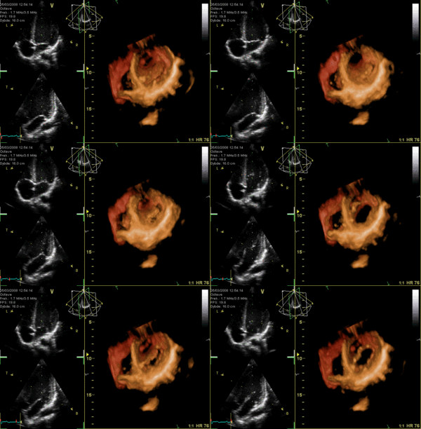
Human Heart Valves – Anatomy, Helpful 3D Animation
By Adam Pick on June 30, 2008
This is very interesting!!! (Especially, for those of you wanting to learn more about human heart valve anatomy.)
I just came across a three dimensional, reconstructive image of the human heart valves. While this animation also shows echocardiogram images (on the left), I was more fascinated with the colorful, 3D presentation of the heart valves opening-and-closing (on the right).
If you look close at this 3D image of the heart, you can see the leaflets of the mitral valve, the aortic valve and the tricuspid valve. Unfortunately, the pulmonary valve is not visible.

So you know…
When I learned that I needed aortic and pulmonary valve replacement operation, I wanted to learn as much as possible about the human heart and its heart valves. My hope is that these types of pictures will help future patients and their caregivers better understand the anatomy of their own hearts before heart valve repair or heart valve replacement operations.
Keep on tickin!
Adam
|
Skyler Salazar says on July 14th, 2009 at 8:09 pm |
|
My 8 year old son, who was born with tetralogy of fallot, has undergone 2 surgeries already…once at 3 months of age and once at 6 years…the first time in the US at Ochsner Clinic in LA and the second time in San Jose Costa Rica…he got a horrible pneumonia in october of last year and was inthe hospital until January….while in the hospital he contracted a bacteria…streptococcus….and it ate the entire repair they had done two years ago….now he is in need of another valve….the drs. want to put a mechanical valve in……what can be done….will he be ok….what do i do for him….help me please…. |
 |
|
Sandra Anderson says on August 30th, 2009 at 8:57 pm |
|
Dear Skyler, I can simpathise with what you’re going through. My 23 year old son was also a “blue baby” and has undergone four surgeries. As a psychiatrist, I worked or many years, helping children and parents of children who were undergoing life threatening procedures. What your son had is known as rheumatic fever, bacteria eating up the repaired heart tissue and often causing a high risk of trombosis (a fragment of cardiac tissue breaking off and entering the bloodstream). In the case of a Tet repair, it is the pulmonary not the aorta which has been replaced, meaning this tissue would end up in the lungs rather than the brain. If the endocarditis is encapsulated, your son should be treated with a high dose of antibiotics as well as a blood thinner, in order to dissolve any fragment that could potentially break appart. I don’t know, in your sons case, if the bacteria has been controlled yet, if encapsulated it could be quite stubborn and resistant to the antibiotic tratment, often taking months months of hospitalization. Normally, surgeons will not proceed until the infection clears out entirely, in order to prevent the risk of it spreading to other organs of the patient’s body. In his particular case, the doctors my opt for a mechanical valve to prevent the infection from reocurring. Once a patient has had rheumatic fever, the person becomes very susceptible to acquiring it again. |
 |


