“Did You Get An M.R.I. Before Heart Valve Surgery?” Asks Gregg
Written By: Adam Pick, Patient Advocate, Author & Website Founder
Published: May 9, 2025
Through your stories and my ongoing research, I have learned that properly diagnosing heart valve disease can be tricky. That said, I always encourage patients to get a second opinion prior to any surgical treatment (if possible).
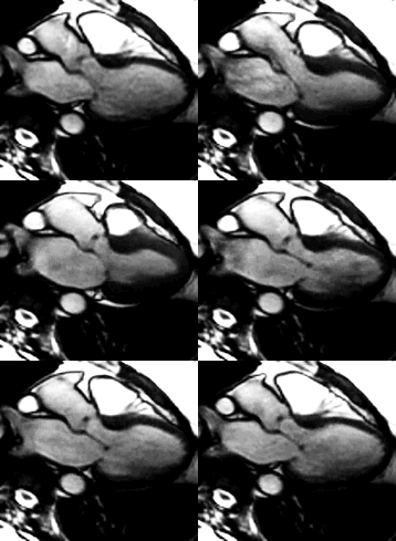
However, Gregg just sent me an interesting question about the use of M.R.I.’s as a diagnostic tool for valvular disorders. [If you have never seen an M.R.I. of the human heart before, please see below… Can you identify the two valves that are opening-and-closing in this image?]

M.R.I. of the Human Heart
Gregg writes, “Hi Adam – Having been diagnosed with a bicuspid valve and an enlarged aorta, I am scheduled for a follow-up echocardiogram this month to determine if there have been any changes to my current condition. My question to you is whether an echo is okay or should I get an MRI? I ask this because during a consult with a surgeon – who is well known for his work in valve repair and valve replacement – he recommended an M.R.I. as a more thorough test. I am curious to know what you and your readers think? Thanks. Gregg”
In my experience, the use of an echocardiogram remains the non-invasive, industry standard for diagnosing heart valve diseases like aortic stenosis and mitral regurgitation. Furthermore, my gut tells me that echocardiogram machines which feature 3D technology will continue to preserve the widespread utility of echocardiograms as the non-invasive benchmark for valve disorder detection and monitoring.
However, I am aware of M.R.I. use as an alternative test for collecting additional information about heart valve disorders.
In fact, the National Heart and Lung Institute suggests, “Cardiac M.R.I. uses a powerful magnet and radio waves to make detailed images of your heart. A cardiac M.R.I. image can confirm information about valve defects or provide more detailed analysis. This information can help your doctor plan your treatment. An MRI also may be done before heart valve surgery to help your surgeon plan for the surgery.”
So you know… Prior to my heart valve surgery, I did not have an M.R.I. performed. Instead, my cardiologists used two distinct echocardiograms to formally diagnose the severe stenosis of my aortic valve.
Related Links:
- Medical Technology Update: See a 3D Echocardiogram Demonstration
- Cardiac Imaging Alert: 4D Flow MRI for Bicuspid Aortic Valve and Aneurysm Risk Analysis
Did you have an M.R.I. as part of your diagnostic process? If so, please click leave a comment below. In advance, thanks for sharing your experience with all of us!!!
Keep on tickin!
Adam
|
jerry says on August 23rd, 2009 at 7:51 pm |
|
It was about 25 years between first diagnosis in college and surgery, and while the first were b&w echos, the latter diagnoses were via color doppler echos with a twist of lemon and an MRI. I can’t say which the cardiologist found most useful, the MRI required I take some metoprolol in order to get my heart rate down to the 60bpm that was the upper limit of the MRI. All of it was covered by insurance — I don’t know the actual costs involved. In the end, my cardiologist seems to really favor an angiogram as the ultimate diagnostic tool. |
 |
|
Lisa says on August 23rd, 2009 at 9:01 pm |
|
I had a standard echocardiogram, a transesophogeal echo, and a specialized cardiac CT scan. With the CT scan there was enough info that I did not have to have an angiogram prior to mitral valve repair surgery. However, I am young and very active so the docs were not worried about any coronary artery disease. As mentioned previously, I had to have I.V. metoprolol as well as sublingual nitroglycerine administered to maximize the images. |
 |
|
Adam Pick says on August 23rd, 2009 at 10:45 pm |
|
Lisa, Like you, I did not require an angiogram prior to surgery given my age and active lifestyle. However, it was very interesting… My first cardiologist, Dr. Bad Bedside Manner, put me on the super, super, super fast track for an angiogram two days later. After reading books, like Coronary, that was another signal (for me at least) that I should consider other cardiologists quickly. I’m glad I did not stick with that Dr. Bad Bedside Manner. I later learned he did the exact same thing to another patient. Thanks for bringing this point up! If any of you are interested in learning more about angiograms / cardiac catheterizations, here is a great patient story: Keep on tickin! Adam |
 |
|
LaVonne Neff says on August 24th, 2009 at 1:38 pm |
|
About the MRI (or MRA: magnetic resonance angiogram): I’m wondering if that would be for the enlarged aorta (aka aneurysm) rather than for the valve problem. I believe CT or MRA scans are more accurate than echocardiograms in calculating the aorta’s diameter. |
 |
|
Rhena Smith says on August 24th, 2009 at 1:59 pm |
|
I just had a heart catheterization on the 7th of August, and in my follow-up with my cardiologist, I asked about an MRI if that could tell more about the situation – particularly I was trying to find out if my aortic valve was bi-cuspid as they were still not sure. I was concerned about other connective tissue disorders that seem to often accompany bi-cuspid AV. Anyway, long story short – they do not routinely doe MRI’s for heart Vavle. They said the gold standard was a hearth cath – which is of course a more invasive procedure than an MRI. |
 |
|
Gail says on August 24th, 2009 at 6:32 pm |
|
My husband had an MRI done pre-op. Initially it was not part of the plan. The cardiologist following him was away on vacation and the doctor we saw thought the valve didn’t look so bad. They decided that the decision would be made to continue with surgery dependent on the MRI results. The MRI showed it needed to be done |
 |
|
nerida says on August 24th, 2009 at 7:21 pm |
|
Gregg, |
 |
|
Chaz says on August 24th, 2009 at 8:33 pm |
|
My surgeon had a CT scan done prior to by aortic valve replacement. He was concerned that the leakage/tear might be larger than indicated by the echocardiagrm and he wanted to prepare for other surgical “possibilities” with more exact measurments. I do know that he used the CD of the CT scan during the operation |
 |
|
MJ Samer says on August 25th, 2009 at 11:04 am |
|
I had a cardiac MRI last week at Northwestern Memorial Hospital in Chicago which my cardiologist told me gave them much clearer data than just an echocardiogram. I will need pulmonary valve replacement and probably tricuspid valve replacement, too in the next couple of months. They want to do one more diagnostic test: a cardiac catheterization. |
 |
|
SNBolding says on October 15th, 2013 at 5:36 pm |
|
My husband had bypass surgery and (mechanical) aortic valve replacement on 10/3. He was comatose for several days, now he is awake but unable to move. The Neurologist wants to do an MRI to help determine the cause of paralysis; however I’m told MRIs cannot be done on a patient with a mechanical valve, at least not in the near future. Does anyone have any information on MRIs after AVS? |
 |


