Surgeon Q&A: “Is it Possible to Remove the Calcium From My Bicuspid Aortic Valve?” asks Rachel
By Adam Pick on February 10, 2015
I just received a great question from Rachel about calcium build-up on bicuspid aortic valves.
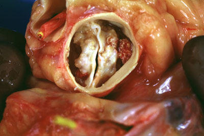
Rachel writes to me, “Hi Adam, At 49, I was recently diagnosed with a severely calcified bicuspid aortic valve. I’m told that I will need surgery in the next year. I don’t really have any symptoms… yet. My question is about the calcium on the leaflets. Is it possible for the surgeon to simply remove the calcium to improve the functioning of the valve? Or, does the calcium always damage the valve leaflets to the point where it needs to be replaced? Thanks, Rachel.”

To provide Rachel an expert response, I reached out to Dr. Glenn Barnhart at the Swedish Heart Institute in Seattle, Washington. So you know, Dr. Barnhart has performed over 2,000 heart valve procedures during his career.
In our community, Dr. Barnhart has successfully treated many patients including Mark Strayhan, Ernest Gee, Marjorie Peterson, Michael Marthaller and Robert Fairchild.

Dr. Glenn Barnhart – Heart Surgeon
In his response to Rachel’s question, Dr. Barnhart shared several insights about calcium deposits on bicuspid aortic valves:
Hi Rachel, I will be happy to answer your question, but first let me give some background information. Patients like yourself with bicuspid aortic valve have a calcium build up in the tissues of their aortic valve leaflets over many years. These calcium deposits start with microscopic bits of calcium within the thin layers of the valve tissue. There are several proposed reasons why calcium buildup and deposits occur; there is in some cases actual bone elements within the aortic valve tissue.
Dr. Barnhart then addressed the impact of calcium on the ability of the valve to function properly:
As the calcium buildup occurs this restricts the leaflet mobility and causes the valve to become tight. The result of this increasing tightness is much like a garden hose in which you “pinch”, causing the water shooting out of the garden hose to increase it’s velocity and come out much faster. This is how the degree of tightness of your aortic valve is measured by your cardiologist with the echocardiogram. We know that the nature of this progression of calcium buildup is to continue to get worse and ultimately cause the patient serious problems including shortness of breath, chest pain, dizziness or congestive heart failure due to back of the blood on the heart and lungs.
Lastly, in his closing remark, Dr. Barnhart discussed the treatment of bicuspid aortic valves with calcium deposits:
Some of the earliest attempts at heart surgery were directed towards patients with problems in their valves. Aortic valve replacement became a standard of care in the 1980’s. In the early and mid-1990’s, surgeons investigated using ultrasound to remove the calcium deposits from aortic valves with the hope of preserving the patient’s native valve. Unfortunately, the majority of these patients returned soon after surgery with holes in their native valve causing them to have significant leakage and some had no significant benefit; these patients had to undergo surgery again to replace the aortic valve. Today, there are several very good options for aortic valve replacement. Talk to your cardiologist and surgeon about the pros and cons of each choice. In many cases, the choice may be based on lifestyle and, in the end may be yours.
Many thanks to Rachel for her questions and a special thanks to Dr. Barnhart for sharing his research and clinical experiences with our patient community.
Keep on tickin!
Adam


