Calcified Aortic Heart Valves: Inside Dr. Doolabh’s Operating Room!
Written By: Adam Pick, Patient Advocate, Author & Website Founder
Published: November 4, 2024
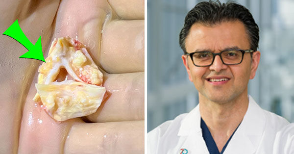
Together, our patient community has a learned a lot about calcified aortic heart valves over the years. As you may know, a calcified heart valve can lead to severe aortic stenosis which is an under-diagnosed and life-threatening form of heart disease. That said, one of the big questions I continue to get from patients is, “What does a calcified aortic valve look like?”
Thanks to Dr. Neelan Doolabh, a leading minimally-invasive heart valve surgeon at UT Southwestern in Dallas, Texas, here are five pictures taken during recent aortic valve replacement surgeries performed by Dr. Doolabh. For example, that is a tri-leaflet aortic valve that has severe calcification all over the leaflets.

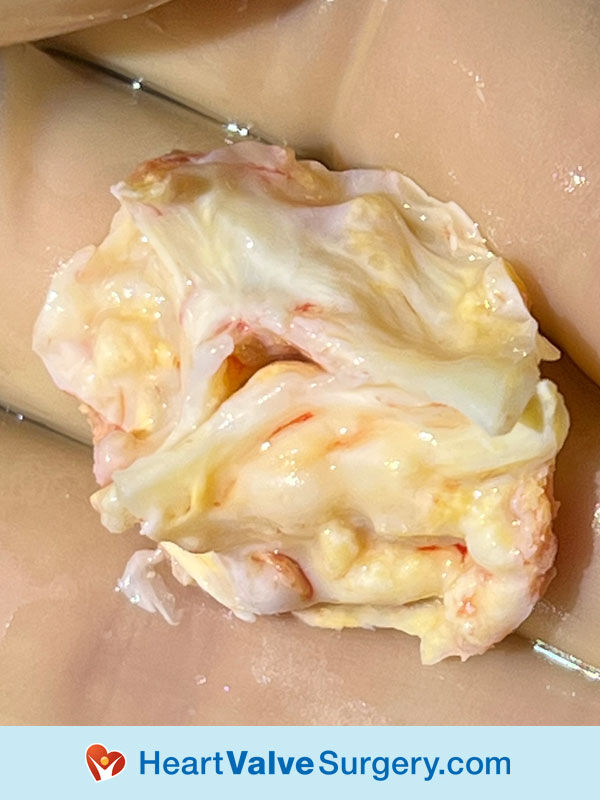
Here is a close-up of another calcified aortic valve that Dr. Doolabh had to replace.

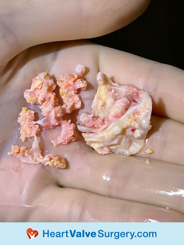
Holy Moly! Here is an extremely calcified aortic valve. You can see all the calcium embedded within the leaflet tissue has essentially broken apart as Dr. Doolabh removed the defective heart valve.

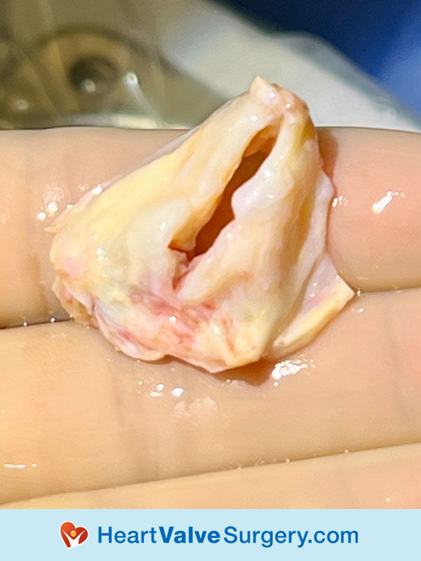
Dr. Doolabh also sent us a calcified bicuspid aortic valve. As you are probably aware, up to 2% of the population has a bicuspid aortic valve which is a congenital heart defect.

Many Thanks Dr. Neelan Doolabh and UT Southwestern!
On behalf of our patient community, thanks so much to Dr. Neelan Doolabh and the entire team at UT Southwestern for taking such great care of our patients with calcified aortic valves.
We really appreciate your support of our educational initiatives!
Related Links:
- Andy Posts First “5-Star Review Video” for Dr. Neelan Doolabh
- Video: Minimally-Invasive Aortic Valve Replacement with Dr. Neelan Doolabh
- See Dr. Doolabh’s Instagram Here!
Keep on tickin!
Adam


